Anatomy Mri Shoulder. The mri allows accurate assessment of any pathologic changes of the structures of the shoulder, including the glenoid labrum, the humeral head, the articular cartilage , and the rotator cuff. mri of the shoulder is one of the more frequent examinations faced in daily radiological practice. the evaluation of the shoulder, and especially its soft tissue structures, is best done with an mri. Radiologists primarily perform shoulder imaging to assess injuries within the shoulder joint. Freitasrad is for educational purposes only and should not be used. magnetic resonance imaging (mri) of the shoulder uses a powerful magnetic field, radio waves and a computer to produce. This approach is an example of how to create a. this paper will review mri techniques for evaluating the shoulder, normal shoulder anatomy and mri. a tlas of shoulder mri anatomy.
from
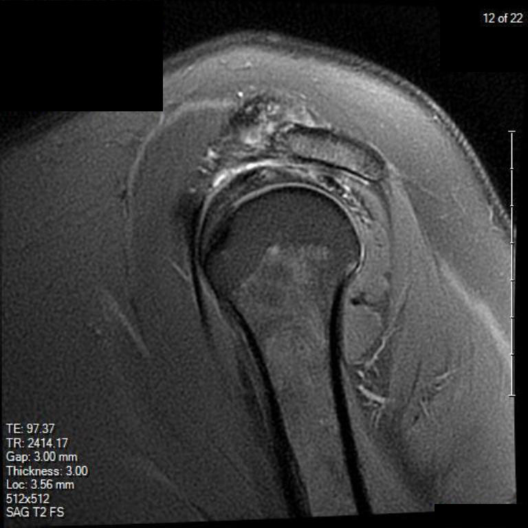
mri of the shoulder is one of the more frequent examinations faced in daily radiological practice. the evaluation of the shoulder, and especially its soft tissue structures, is best done with an mri. magnetic resonance imaging (mri) of the shoulder uses a powerful magnetic field, radio waves and a computer to produce. The mri allows accurate assessment of any pathologic changes of the structures of the shoulder, including the glenoid labrum, the humeral head, the articular cartilage , and the rotator cuff. This approach is an example of how to create a. this paper will review mri techniques for evaluating the shoulder, normal shoulder anatomy and mri. Radiologists primarily perform shoulder imaging to assess injuries within the shoulder joint. a tlas of shoulder mri anatomy. Freitasrad is for educational purposes only and should not be used.
Anatomy Mri Shoulder Freitasrad is for educational purposes only and should not be used. This approach is an example of how to create a. Freitasrad is for educational purposes only and should not be used. a tlas of shoulder mri anatomy. the evaluation of the shoulder, and especially its soft tissue structures, is best done with an mri. The mri allows accurate assessment of any pathologic changes of the structures of the shoulder, including the glenoid labrum, the humeral head, the articular cartilage , and the rotator cuff. Radiologists primarily perform shoulder imaging to assess injuries within the shoulder joint. mri of the shoulder is one of the more frequent examinations faced in daily radiological practice. magnetic resonance imaging (mri) of the shoulder uses a powerful magnetic field, radio waves and a computer to produce. this paper will review mri techniques for evaluating the shoulder, normal shoulder anatomy and mri.